
Don’t become VOMIT
Not literally VOMIT, but a Victim Of Medical Imaging Technology.
Please read and then share to your friends and family. The United States is ranked number 1 in health care costs yet consistency ranks as one of the worst. I believe over imaging is certainly one of the biggest contributors.
Taken from the infograph published by the APTEI (Advanced Physical Therapy Education Institute):
- Medical imaging procedures such X-rays, ultrasounds, CT Scans and MRIs can be very valuable for identifying serious medical conditions such as fractures, dislocations, spinal cord injuries, etc. However, once serious problems have been ruled out by a doctor, minor findings are of no value in helping to explaining the far majoring of aches and pains.
- In fact, not only are the majority of imaging results not helpful, studies show that they are even harmful from a psychological point of view. Especially when reports tell patients they have arthritis, degenerative discs, disc budges, tendon tears, etc.
- Everyday, thousands of patients across Canada (I would obviously include the United States), become a VOMIT (Victim Of Medical Imaging Technology). Those who have become a VOMIT have been shown to have more doctor’s visits, longer lasting pain, more disability and a lower sense of well-being (1,2,3).
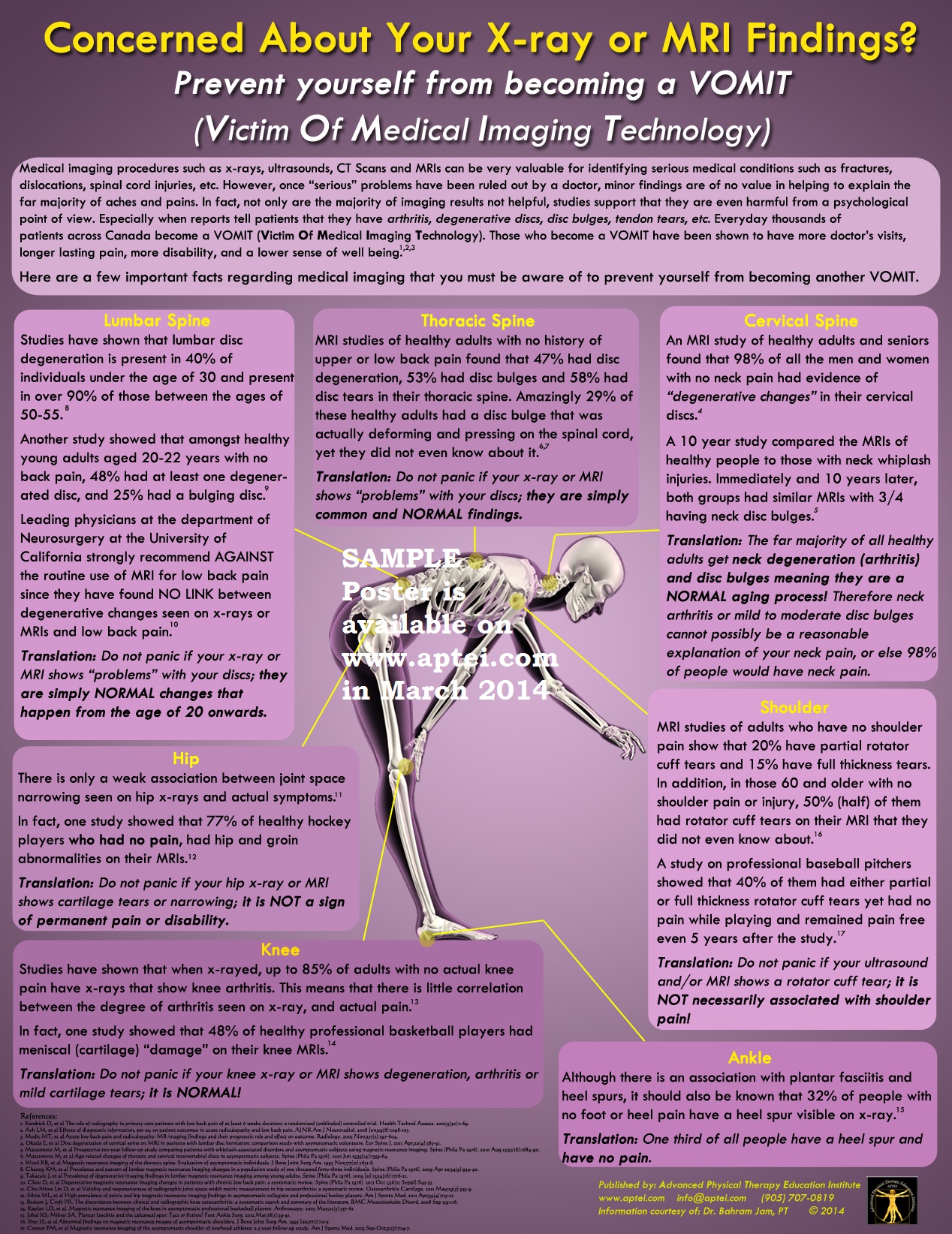
Here are a few important facts regarding medical imaging that you must be aware of to prevent yourself from being another VOMIT:
- Lumbar spine
- Studies have shown that lumbar disc degeneration is present in 40% of individuals under the age of 30 and present in 90% of those between the ages of 50-55 (8).
- Another study showed that amongst healthy adults ages 20-22 years with no back pain, 48% had at least one degenerative disc and and 25% had a bulging disc (9)
- Leading physicians at the department of Neurosurgery and the University of California strongly recommend AGAINST the routine use of MRI for low back pain since they found NO LINK between degenerative changes seen on x-rays or MRIs and low back pain (10).
- Translation: Do not panic if your x-ray or MRI shows “problems” with your discs, they are simply normal changes that happen from the age of 20 onwards.
- Cervical spine
- An MRI study of healthy adults and seniors found that 98% of all the men and women with no neck pain had “degenerative changes” in their cervical discs (4).
- A 10 year study compared the MRIs of healthy people to those with neck whiplash injuries. Immediately and 10 years later, both groups had similar MRIs with 3/4 having neck disc bulges (5).
- Translation: The far majority of all healthy adults get neck degeneration (arthritis) and disc bulges meaning they are NORMAL aging process! Therefore neck arthritis or mild to moderate disc bulges cannot possibly be a reasonable explanation for your neck pain, or else 98% of people would have neck pain.
- Thoracic spine
- MRI studies of healthy individuals with no history of upper or lower back pain found that 47% had disc degeneration, 53% had disc bulges, and 58% had disc tears in their thoracic spine.
- Amazingly 29% of those healthy adults have a disc bulge that was actually deforming and pressing on the spinal cord, yet they did not know about it (6,7).
- Translation: Do not panic if your x-rays or MRI shows “problems” with your discs, they are simply common and normal findings.
- Hip
- There is only a weak association between joint space narrowing seen on x-rays and actual symptoms (11).
- In fact, one study showed that 77% of healthy hockey players who had no pain, had hip and groin abnormalities on their MRIs (12).
- Translation: Do not panic if your hip x-ray or MRI shows cartilage tears or narrowing, it is not a sign of permanent pain or disability.
- Knee
- Studies have shown that when x-rayed, up to 85% of adults with no actual knee pain have x-rays that show knee arthritis. This means that there is little correlation between degree of knee arthritis, and actual pain (13).
- In fact, one study showed that 48% of healthy professional basketball players had meniscal (cartilage) damage on their MRIs (14).
- Translation: Do not panic if your knee x-ray or MRI shows degeneration, arthritis, or mild cartilage tears, it is NORMAL.
- Ankle
- Although there is an association between plantar fasciitis and heel spurs, it should also be known that 32% of people with no foot or heel pain have a heel spur visible on x-ray (15).
- Translation: 1/3 of all people have a heel spur and have no pain.
- Shoulder
- MRI studies of adults who have no shoulder pain show that 20% have partial rotation cuff tears and 15% have full thickness tears. In addition, of those 60 and older with no shoulder pain or injury, 50% (half) of them had rotator cuff tears on their MRI that they did not know about (16).
- A study on professional baseball pitchers showed that 40% of them had either partial or full thickness rotator cuff tears yet had no pain while playing and remained pain free even 5 years after the study (17).
- Translation: Do not panic if your ultrasound and/or MRI shows a rotator cuff tear, it is not necessarily associated with shoulder pain.
I do not have a list of the sources, they are contained in the infograph. The VOMIT poster is available from the Advanced Physical Therapy Education Institute website for $20
I am not responsible for this info, I am just merely sharing what I have found. This information belongs to the APTEI (Advanced Physical Therapy Education Institute).